
Abstract
PURPOSE
Ultrasound (US)-guided marker placement is an emerging method for sentinel lymph node biopsy (SLNB), and this study aimed to investigate the concordance between US-guided marker placement for peri-foraminal lymph nodes (LNs) in the axillary cribriform fascia and blue dye-stained SLNs, especially the utility of the two concurrent labeling methods for SLNB in patients with early-stage breast cancer.
METHODS
Female patients with clinically staged T1-2N0M0 breast cancer were enrolled. Patients received US-guided marker placement on the day before surgery, followed by blue dye staining. SLN biopsy was performed on the blue dye-stained LNs first and then on the US-guided markers.
RESULTS
A total of 96 eligible patients were enrolled. The detection rates of US-guided localization, blue dye, and their combination were 100.0%, 95.8%, and 100.0%, respectively. Among the 92 patients with blue dye staining, US-guided markers were found in blue-stained LNs from 91 patients (concordant) but failed in 1 patient (non-concordant); among the 4 patients without blue-stained LNs, US-guided markers were found in all 4 patients (non-concordant), resulting in a general concordance rate of 94.8% (91/96). Notably, among the 92 patients with blue dye staining, 23 had LN metastasis diagnosed through SLNB, whereas among the 4 patients without blue-stained LNs, the SLNB was performed based on US-guided markers, which revealed 3 patients with LN metastasis and 1 without. Moreover, combining the two labeling methods concurrently identified 26 patients with LN metastasis.
CONCLUSION
US-guided localization combined with blue dye concurrently may be a satisfied option for SLNB detection.
CLINICAL SIGNIFICANCE
US-guided localization combined with blue dye offers an accessible, anatomically grounded, and radiation-free strategy for optimizing the SLNB procedure, especially in settings where radioactive isotopes are not available.
Keywords: Breast cancer, ultrasound-guided marker placement, axillary cribriform fascia, blue dye, sentinel lymph node biopsy
Main points
• Ultrasound (US)-guided marker placement demonstrated high concordance with blue dye for sentinel lymph node biopsy (SLNB) in breast cancer.
• US-guided marker placement combined with blue dye achieved a satisfied detection rate of SLNB in breast cancer.
• Collectively, US-guided localization plus blue dye may be an accessible, anatomically grounded, and radiation-free strategy for optimizing SLNB in breast cancer.
Breast cancer remains the most prevalent cancer in women, especially those aged between 40 and 49 years, with approximately 2.31 million new cases and 0.67 million deaths every year worldwide, seriously threatening women’s physical and mental health., Surgical treatment is a fundamental component of the comprehensive management of breast cancer, with axillary lymph node dissection (ALND) enabling accurate LN staging, removing potential metastatic LNs, achieving superior local recurrence control, and improving the survival of patients.,, However, ALND is associated with several complications, such as upper lymphedema, shoulder dysfunction, paresthesia, brachial plexus injury, and wound infection, which seriously affect patients’ recovery and quality of life.,,
With improvements in breast cancer screening, an increasing number of patients with breast cancer are being diagnosed with clinically negative axillary LNs at an early stage., In this context, the clinical utility of sentinel LN biopsy (SLNB) has become prominent, as the first node or group of nodes receiving lymphatic drainage from the primary tumor can be used to estimate the pathological status of all axillary LNs and serve as a key predictor of recurrence risk and overall survival in patients with breast cancer., Due to its high accuracy, the status of the SLN is critical for decisions regarding both the type of surgery and related treatments., Moreover, SLNB is associated with less trauma and fewer complications and is widely applied in patients with early-stage breast cancer with clinically negative axillary LNs to avoid unnecessary ALND.,
SLN tracing methods mainly include the blue dye method (such as methylene blue, patent blue, and isosulfan blue), the nuclide method combined with the dye method, and the fluorescence tracer method.,, The nuclide method (e.g., 99ᵐTc) offers high detection rates but with radiation exposure, logistical complexity, and limited accessibility in many institutions. The fluorescence method (e.g., indocyanine green) provides real-time visualization but requires specialized equipment and has limited tissue penetration. By contrast, the blue dye method is simple, intuitive, and widely available, yet its detection rate (approximately 91%–95%) and false-negative rate remain suboptimal. These comparative limitations contextualize the rationale for developing an accessible, radiation-free adjunct, such as ultrasound (US)-guided marker placement, to complement the blue dye and improve SLNB accuracy, particularly in resource-limited settings where nuclide and fluorescence techniques are unavailable.,, However, the blue dye method still has the disadvantages of a relatively low detection rate and high false-negative rate; therefore, the exploration of better methods for SLN tracing and subsequent SLNB remains vital.
US-guided marker placement is a technique that is being increasingly explored for the precise localization of breast lesions and LNs. It facilitates accurate surgical excision and allows for the effective tracking of tumor response throughout neoadjuvant chemotherapy., Notably, US-guided marker placement is simple, stable, and safe for axillary LNs in patients with breast cancer. Moreover, our previous study revealed that SLNB via the axillary cribriform fascia (ACF) with blue dye anatomically covering the SLN area improved intraoperative blood loss, drainage flow, and upper limb dysfunction. The ACF is formed by the fusion of superficial and deep fascial layers, creating natural foramina that serve as obligate passageways for breast lymphatic drainage into the axilla; consequently, LNs situated adjacent to these foramina represent the first echelon of nodal filtering, functionally defining them as SLNs., This anatomical framework provides the logical basis for US-guided targeting of peri-foraminal LNs as surrogates for true SLNs. Thus, we hypothesized that the US-guided identification of peri-foraminal LNs in the ACF may represent SLNs, but related studies are rare.
This study aimed to investigate whether US-guided marker placement of peri-foraminal LNs in the ACF is concordant with blue dye-stained SLNs and to determine whether combining these two methods improves SLNB performance in patients with early-stage breast cancer.
Methods
Patients
From May 2023 to July 2024, female patients with clinically staged T1-2N0M0 invasive breast cancer who were scheduled to undergo SLNB were prospectively enrolled from the Department of Breast Surgery, Tangshan People’s Hospital, China. The inclusion criteria were as follows: (1) aged ≥ 18 years; (2) unifocal, primary breast cancer confirmed by core needle biopsy; and (3) no clinical or imaging evidence of axillary LN metastasis from US. Patients with prior axillary surgery, neoadjuvant chemotherapy, or systemic metastases were excluded. This study was approved by the Ethics Committee of Tangshan People’s Hospital with approval number RMYY-LLKS-2024023 and approval date 5/24/2024. Moreover, this study was registered on the Chinese Clinical Trial Registry (https://www.chictr.org.cn) with the access number ChiCTR2400087357. In addition, all patients provided written informed consent.
Ultrasound-guided marker placement
US examination was performed using a Philips EPIQ 7 ultrasound system (Philips Healthcare, Bothell, WA, USA) equipped with a high-resolution linear transducer (eL18-4 MHz). US-guided marker placement in peri-foraminal LNs in the ACF was performed using an UltraClip™ breast tissue marker needle with a US-enhanced coil (Bard, USA). In brief, on the day prior to surgery, the patients underwent high-resolution US examination of the axilla. Attention was given to the cribriform foramen area adjacent to the mammary gland in the affected axilla, particularly the ACF, cribriform foramen, and LNs; then, the LN closest to the mammary gland and nearest to the ACF foramen was identified (-). A sterile 18G marker needle was subsequently inserted into the cortical margin of the identified LN under real-time US guidance, carefully avoiding the adjacent vascular structures (-). The needle position and corresponding US images were documented, and the skin overlying the insertion site was marked to guide intraoperative identification.
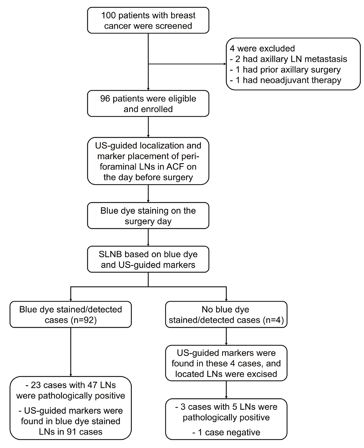
Study flowchart illustrating patient enrollment and procedures. SLNB, sentinel lymph node biopsy; US, ultrasound; LN, lymph node; ACF, axillary cribriform fascia.
Blue dye
On the day of surgery, 2–3 mL of 1% methylene blue dye was injected subcutaneously into the periareolar region and around the tumor of the affected breast approximately 15 minutes before the skin incision.
Sentinel lymph node biopsy procedure
A standard SLNB procedure was performed through a small axillary incision based on the blue dye and US-guided markers. First, blue dye-stained LNs were carefully identified and excised (-). Second, the surgeon explored the region corresponding to the US-guided marker placement, and all visible LNs (not only the marker-localized ones) within this anatomically defined area were retrieved, regardless of the presence of dye staining.
Third, visual observation and radiographic examination were performed that confirmed the presence of the US-guided marker in the excised LN specimens (-). Finally, all excised LNs were subjected to intraoperative frozen section analysis and routine histopathological examination.
Sample size calculation
The minimum sample size was calculated on the basis of the following settings: a paired design, a blue dye detection rate estimated at 90%, a US-guided marker placement detection rate estimated at 100%, power of 90%, and α: 0.05 (two tailed), resulting in a minimum sample size of 89 cases. This study included 96 patients, which exceeded the minimum sample size.
Data collection and outcome measures
Patient characteristics, including age, tumor size, clinical TNM stage, tumor location, and molecular subtypes, were recorded. The primary outcome was the concordance rate between US-marked LNs in the ACF area and blue dye-stained SLNs. The secondary outcomes included the number of detected LNs, number of detected cases, detection rate (by cases), average number of LNs per case, and SLNB pathological findings. Detection rate (%): (number of detected cases/all cases) × 100%.
Statistical analysis
For statistical analysis, SPSS version 26.0 (IBM Corp., Armonk, NY, USA) was used. Data are presented as the mean ± standard deviation, n, %, or n (%). Comparisons were performed using the chi-square test, Fisher’s exact test, Student’s t test, or McNemar test, as appropriate. A two-sided P value < 0.05 was considered statistically significant.
Results
Study flow
A total of 100 patients with breast cancer were initially screened, with 2 excluded because they had axillary LN metastasis, 1 because of prior axillary surgery, and 1 because of neoadjuvant therapy. The remaining 96 patients were eligible and included in this study. The patients underwent US-guided localization and marker placement of peri-foraminal LNs in ACF on the day before surgery. On the day of surgery, blue dye staining was performed, and SLNB was conducted first on the blue dye-stained LNs and then on the US-guided markers ().
Patient characteristics
The mean age of the included patients was 41.4 ± 7.9 years, with a mean tumor size of 1.3 ± 0.3 cm (). Thirty-nine (40.6%) and 57 (59.4%) patients had clinical TNM stage I and IIA disease, respectively. Fifty-nine (61.5%), 13 (13.5%), 14 (14.6%), and 10 (10.4%) patients had tumors located in the upper outer quadrant, lower outer quadrant, upper inner quadrant, and lower inner quadrant, respectively. Moreover, 30 (31.2%), 33 (34.4%), 21 (21.9%), and 12 (12.5%) patients were classified as luminal A, luminal B, human epidermal growth factor receptor 2 positive, and triple negative, respectively.
Table 1. Patient characteristics
Parameters | Patients with breast cancer (n = 96) |
|---|---|
Age (years), mean ± SD | 41.4 ± 7.9 |
Tumor size (cm), mean ± SD | 1.3 ± 0.3 |
Clinical TNM stage, n (%) | |
I | 39 (40.6) |
IIA | 57 (59.4) |
Tumor location, n (%) | |
Upper outer quadrant | 59 (61.5) |
Lower outer quadrant | 13 (13.5) |
Upper inner quadrant | 14 (14.6) |
Lower inner quadrant | 10 (10.4) |
Molecular subtype, n (%) | |
Luminal A | 30 (31.2) |
Luminal B | 33 (34.4) |
HER2 positive | 21 (21.9) |
Triple negative | 12 (12.5) |
SD, standard deviation; TNM, tumor-node, and metastasis; HER2, human epidermal growth factor receptor 2.
Comparison of detection rates
A total of 293 LNs in 96 patients were detected by US-guided localization, with a detection rate of 100.0%, whereas a total of 282 LNs in 92 patients were detected by blue dye staining, resulting in a detection rate of 95.8% (). Therefore, the detection rate was not significantly different between US-guided localization and blue dye staining (P = 0.125). Furthermore, the average number of LNs detected per patient was 3.05 ± 0.49 by US-guided localization and 3.07 ± 0.46 by blue dye staining, with no significant difference between the two methods (P = 0.851). Notably, when the two labeling methods (blue dye + US) were combined, the detection rate reached 100.0%, with an average number of LNs detected per case of 3.06 ± 0.52.
Table 2. Lymph node detection using ultrasound and blue dye
Parameters | Number of detected LNs | Number of detected cases | Detection rate by cases | P1 value | Average LNs per case | P2 value |
US (n = 96) | 293 | 96 | 100.0% | 0.125 | 3.05 ± 0.49 | 0.851 |
Blue dye (n = 96) | 282 | 92 | 95.8% | - | 3.07 ± 0.46 | - |
Blue dye + US | 294 | 96 | 100.0% | − | 3.06 ± 0.52 | − |
P1 represented the comparison of detection rates between US and blue dye; P2 represented the comparison of average LNs per case between US and blue dye. LN, lymph node; US, ultrasound.
Concordance between ultrasound-guided localization and blue dye staining
In the 92 patients with blue dye staining, US-guided markers were found in blue dye-stained LNs from 91 patients (concordant) but failed in 1 patient (non-concordant). In the 4 patients without blue dye staining, US-guided markers were found in all 4 (non-concordant). Taken together, the concordance rate between US-guided localization and blue dye staining was 94.8% ().
Table 3. Concordance rate between ultrasound and blue dye detection
Parameters | In blue dye-detected cases | Concordance rate | ||
Yes (n = 92) | No (n = 4) | |||
Ultrasound detection | Concordant | 91 | 0 | 94.8% |
Non-concordant | 1 | 4 | ||
Pathological findings from sentinel lymph node biopsy
In the 92 patients with blue dye staining, 23 had positive LN metastasis revealed by SLNB, consisting of 47 positive LNs. Notably, in the 4 patients with no blue dye staining, a sum of 11 LNs were excised based on the sites of the US-guided markers and proposed for biopsy, revealing 3 patients with positive LN metastasis (consisting of 5 positive LNs) and 1 patient with negative LN metastasis. This indicated that US-guided localization improved the accuracy of SLNB. Moreover, as a result of LN identification using the blue dye combined with US-guided localization method, 26 patients were pathologically confirmed through SLNB to have positive LN metastasis, resulting in the identification of 52 positive LNs (). In addition, among the 91 concordant cases, 22 patients had positive LN metastasis (consisting of 45 positive LNs).
Table 4. Pathological findings from SLNB
Parameters | SLNB positivity by LNs | SLNB positivity by cases |
|---|---|---|
Blue dye detected (92 cases) | 47 | 23 |
Blue dye non-detected but US detected (4 cases) | 5 | 3 |
US detected (96 cases) | 50 | 25 |
Blue dye + US detected (96 cases) | 52 | 26 |
SLNB, sentinel lymph node biopsy; US, ultrasound; LN, lymph node. | ||
Safety profile of ultrasound-guided marker placement
In general, no adverse events or complications directly related to the US-guided marker placement procedure were observed. Moreover, no marker dislodgement, migration beyond the LN capsule, or infections were observed.
Discussion
Currently, the need for ALND in patients with early-stage breast cancer with negative axillary LNs remains a subject of debate.,, Previous studies have indicated that patients with negative SLNs may be exempt from ALND, suggesting that SLNB can effectively serve as a substitute for ALND for early-stage breast cancer.,, Moreover, SLNB has been established as a safe, straightforward, and reliable technique that helps patients avoid a range of complications associated with ALND.
A comprehensive meta-analysis revealed that blue dye is a standard labeling method for SLNB, with a detection rate of approximately 91% (95% CI: 88%–94%) in patients with breast cancer. Moreover, combining it with another labeling method concurrently further improved the detection rate of blue dye for SLNB., For instance, indocyanine green and blue dye concurrently achieve a detection rate of 98.5%, and Tc99-labeled sulfur colloid plus blue dye concurrently achieves a detection rate of 96.4%–100.0% for SLNB in patients with breast cancer. US-guided localization has also been applied to SLNB in breast cancer, with a detection rate of 96.3%. However, investigations that combine US-guided marker placement and blue dye methods lack sufficient evidence. In the present study, US-guided localization, blue dye-guided staining, and their combination achieved detection rates of 100.0%, 95.8%, and 100.0%, respectively, for SLNB in patients with early-stage breast cancer, indicating that US-guided localization improves the detection rate of blue dye.
Importantly, this study focused on assessing the concordance between US-guided localization of peri-foraminal LNs in the ACF and traditional blue dye-stained SLNs in patients with early-stage breast cancer and revealed an exceptionally high concordance rate of 94.8%, indicating that the peri-foraminal LNs in the ACF visualized under US are highly representative of true SLNs. This strong overlap supports the anatomical reliability of US-guided localization based on axillary fascial structures. Anatomically, the peri-foraminal region in the ACF corresponds to a critical convergence area for breast lymphatic drainage. The ability of US to visualize these structures and target LNs within or adjacent to the foramen provides a solid anatomical basis for its use in SLN identification.,
Although the concordance was high, one patient exhibited “skip metastasis” in this study, where the metastatic SLN lay outside the US-visible peri-foraminal region in the ACF. Although rare, such conditions have been reported previously., Skip metastasis, which is commonly recognized as the metastatic involvement of higher-level axillary nodes without level I nodes, occurs in approximately 1.5%–5.3% of breast cancer cases. Within the ACF-based anatomical framework, several mechanisms may explain this phenomenon. First, accessory lymphatic channels may bypass the peri-foraminal region entirely, traversing the ACF by non-sieve routes., Second, tumor-induced lymphatic obstruction can reroute flow into collateral pathways that circumvent typical first-echelon nodes. Third, interindividual variations in the number and distribution of cribriform foramina may result in incomplete lymphatic capture by a single peri-foraminal node. Thus, although US-guided marker placement is highly accurate, it should be complemented by intraoperative exploration, particularly in anatomically variant cases.
In this study, among the 92 patients with blue dye staining, 23 were confirmed by SLNB to have LN metastasis; notably, among the 4 patients who did not have blue dye staining, LNs were excised on the basis of US-stained sites, after which 3 patients were confirmed by SLNB to have LN metastasis and 1 was negative. These findings suggest that US can reliably identify metastatic SLNs when the blue dye method fails, particularly in patients who are elderly or obese, thereby reducing the risk of false negatives and supporting more precise surgical decision-making.
Beyond clinical performance, the cost profile of US-guided marker placement combined with blue dye warrants consideration, particularly when compared with alternative dual-tracing methods for SLNB. The most widely established dual-tracing approach in many high-resource settings is the combination of radioactive colloid (e.g., 99ᵐTc-labeled sulfur colloid) with blue dye. However, this method incurs substantial costs related to nuclear medicine facilities, radiopharmaceutical preparation, gamma probe maintenance, and personnel training for radiation handling and waste disposal., In resource-limited or non-specialized centers where such infrastructure is unavailable, the isotope-based dual tracing may not be feasible. Another increasingly used dual modality is indocyanine green combined with blue dye, which eliminates radiation but requires a near-infrared fluorescence imaging system and dedicated cameras, representing a substantial capital investment and recurrent maintenance cost., Regarding the US-guided marker placement, its cost mainly lies in a standard high-resolution US machine (commonly already present in the institution) and a disposable marker needle, whose combination with blue dye was less costly than radioactive colloid combined with blue dye and indocyanine green combined with blue dye. Specifically, the cost of US-guided marker placement combined with blue dye is approximately Chinese Yuan (CNY) 600 in our institution, whereas that of radioactive colloid combined with blue dye is approximately CNY 1,200 and that of indocyanine green combined with blue dye is approximately CNY 1,400.
Despite the clinical and cost advantages discussed above, it is important to acknowledge a disadvantage—US-guided marker placement requires a pre-operative procedure with US clipping on the day before surgery. This entails an additional hospital visit, a second needle insertion under US guidance, and the placement of one or more metallic markers into axillary LNs.
Several limitations of this study should be mentioned. First, as a single-center study with a moderate sample size, the generalizability of the study findings is limited, and future multicenter studies with larger cohorts are warranted. Second, the superficial projection of LNs relative to the ACF may vary with changes in patient positioning between US localization and surgery, introducing potential bias. Third, this study did not employ subcutaneous contrast-enhanced US for LN imaging, which could improve the precision of identifying the primary draining SLN. Fourth, ALND was not performed to clarify the false-negative rate of US-guided localization.
In conclusion, US-guided localization of peri-foraminal LNs in the ACF exhibits a satisfactory detection rate and has high concordance with blue dye-stained SLNs in patients with early-stage breast cancer. Notably, US-guided localization combined with blue dye offers an accessible, anatomically grounded, and radiation-free strategy for optimizing SLNB, particularly in settings where radioactive isotopes are not available. However, further validation with large-scale samples and multicenter studies is still needed.
Footnotes
Conflict of interest disclosure
The authors declared no conflicts of interest.
Funding
This study was supported by the Wu Jieping Medical Foundation Special Research Project Grant (project number: 320.6750.2024-18-36), and the Excellent Going Abroad Experts’ Training Program in Hebei Province (project number: 2023-36).
References
Bray F, Laversanne M, Sung H, et al. Global cancer statistics 2022: GLOBOCAN estimates of incidence and mortality worldwide for 36 cancers in 185 countries. CA Cancer J Clin. 2024;74(3):229-263.
Filho AM, Laversanne M, Ferlay J, et al. The GLOBOCAN 2022 cancer estimates: data sources, methods, and a snapshot of the cancer burden worldwide. Int J Cancer. 2025;156(7):1336-1346.
Bartels SAL, Donker M, Poncet C, et al. Radiotherapy or surgery of the axilla after a positive sentinel node in breast cancer: 10-year results of the randomized controlled EORTC 10981-22023 AMAROS trial. J Clin Oncol. 2023;41(12):2159-2165.
Heidinger M, Weber WP. Axillary surgery for breast cancer in 2024. Cancers (Basel). 2024;16(9):1623.
Weber WP, Hanson SE, Wong DE, et al. Personalizing Locoregional therapy in patients with breast cancer in 2024: tailoring axillary surgery, escalating lymphatic surgery, and implementing evidence-based hypofractionated radiotherapy. Am Soc Clin Oncol Educ Book. 2024;44(3):e438776.
Mattar A, Antonini M, Cavalcante FP, et al. CADONOT: comparing axillary dissection or not in breast cancer surgery. Breast. 2025;81:104453.
Krag DN, Anderson SJ, Julian TB, et al. Sentinel-lymph-node resection compared with conventional axillary-lymph-node dissection in clinically node-negative patients with breast cancer: overall survival findings from the NSABP B-32 randomised phase 3 trial. Lancet Oncol. 2010;11(10):927-933.
Land SR, Kopec JA, Julian TB, et al. Patient-reported outcomes in sentinel node-negative adjuvant breast cancer patients receiving sentinel-node biopsy or axillary dissection: National Surgical Adjuvant Breast and Bowel Project phase III protocol B-32. J Clin Oncol. 2010;28(25):3929-3936. Erratum in: J Clin Oncol. 2010;28(36):5350.
Giaquinto AN, Sung H, Newman LA, et al. Breast cancer statistics 2024. CA Cancer J Clin. 2024;74(6):477-495.
James J, Law M, Sengupta S, Saunders C. Assessment of the axilla in women with early-stage breast cancer undergoing primary surgery: a review. World J Surg Oncol. 2024;22(1):127.
Park KU, Somerfield MR, Anne N, et al. Sentinel lymph node biopsy in early-stage breast cancer: ASCO guideline update. J Clin Oncol. 2025;43(14):1720-1741.
Mamtani A, Barrio AV. Updates in surgical management of the axilla. Oncology (Williston Park). 2025;39(2):70-75.
Gentile D, Tinterri C. Sentinel lymph node biopsy versus axillary lymph node dissection in breast cancer patients undergoing mastectomy. Minerva Surg. 2024;79(5):545-557.
Xia L, Zou Z, Chong L, Wang X, Dong Z, Zhao Y. The value of nanocarbon contrast methylene blue based on dye-based tracer technology in sentinel lymph node biopsy for breast cancer: a systematic review and meta-analysis. PeerJ. 2025;13:e19546.
Rovera G, de Koster EJ, Rufini V, et al. 99mTc-Tilmanocept performance for sentinel node mapping in breast cancer, melanoma, and head and neck cancer: a systematic review and meta-analysis from a European expert panel. Eur J Nucl Med Mol Imaging. 2023;50(11):3375-3389.
Yadav SK, Bharath S, Sharma D, et al. A systematic review and meta-analysis of diagnostic performance of fluorescein-guided sentinel lymph node biopsy in early breast cancer. Breast Cancer Res Treat. 2024;206(1):19-30.
Li J, Chen X, Qi M, Li Y. Sentinel lymph node biopsy mapped with methylene blue dye alone in patients with breast cancer: a systematic review and meta-analysis. PLoS One. 2018;13(9):e0204364.
Li H, Jun Z, Zhi-Cheng G, Xiang Q. Factors that affect the false negative rate of sentinel lymph node mapping with methylene blue dye alone in breast cancer. J Int Med Res. 2019;47(10):4841-4853.
Schulz-Wendtland R, Dankerl P, Dilbat G, et al. Evaluation of newly adapted clip marker system in ultrasound-guided core needle biopsy for suspicion of breast cancer. Geburtshilfe Frauenheilkd. 2013;73(11):1135-1138.
Shaukat S, Haroon J, Afzal F, et al. Role of ultrasound-guided marker placement in breast lesion for precise localization, surgical resection, neoadjuvant chemotherapy response, and post-chemotherapy follow-up. Cureus. 2025;17(8):e89402.
Ji X, Wei M, Wang L, Li J, Gao D, Geng C. Application of ultrasound-guided placement of markers for locating axillary lymph nodes of breast cancer. Gland Surg. 2021;10(11):3067-3074.
Chang JY, Wang W, Shen JL, Zhang Y, Cai HF. Impact of sentinel lymph node biopsy through the axillary cribriform fascia approach on intraoperative indicators and postoperative complications. Updates Surg. 2023;75(3):757-767.
Liu YQ, Zhang XP, Cai HF. Anatomy of the cribriform fascia and its significance in the membranous anatomy of breast surgery. Journal of Regional Anatomy and Operative Surgery. 2025;34(7):634-637.
Dixon JM. Sentinel lymph node biopsy in breast cancer surgery. Ann Surg Oncol. 2016;23(11):3426-3428.
Tejedor L, Gómez-Modet S. Reducing axillary surgery in breast cancer. Cir Esp (Engl Ed). 2024;102(4):220-224.
Manca G, Rubello D, Tardelli E, et al. Sentinel lymph node biopsy in breast cancer: indications, contraindications, and controversies. Clin Nucl Med. 2016;41(2):126-133.
Reimer T, Stachs A, Veselinovic K, et al. Axillary Surgery in breast cancer - primary results of the INSEMA trial. N Engl J Med. 2025;392(11):1051-1064.
Mamtani A, Sevilimedu V, Barrio AV, Morrow M. Axillary dissection is avoidable in most cT1N0 triple-negative & HER2+ breast cancers treated with upfront surgery. Breast Cancer Res Treat. 2025;213(3):355-363.
Yang QH, Zhang XJ. Indocyanine green combined with methylene blue versus methylene blue alone for sentinel lymph node biopsy in breast cancer: a retrospective study. BMC Surg. 2023;23(1):133.
Malhotra C, Pawar R, Patni S, Sucheta, Kaushik M, Sharma N. Efficacy of periareolar versus peritumoral injection of TC99-labelled sulphur colloid and methylene blue dye for detection of sentinel lymph node in patients with early breast cancer: a comparative study. Indian J Surg Oncol. 2021;12(1):119-123.
Cui Q, Dai L, Li J, et al. Contrast-enhanced ultrasound-guided sentinel lymph node biopsy in early-stage breast cancer: a prospective cohort study. World J Surg Oncol. 2023;21(1):143.
Lo C, Lee PC, Yen RF, Huang CS. Most frequent location of the sentinel lymph nodes. Asian J Surg. 2014;37(3):125-129.
Msuya CA, Hartveit F. Skip lesions in the axilla in breast cancer, and their association with micrometastases. Breast Cancer Res Treat. 1991;19(3):277-281.
Canavese G, Catturich A, Vecchio C, et al. Prognostic role of lymph-node level involvement in patients undergoing axillary dissection for breast cancer. Eur J Surg Oncol. 1998;24(2):104-109.
Rosen PP, Lesser ML, Kinne DW, Beattie EJ. Discontinuous or “skip” metastases in breast carcinoma. Analysis of 1228 axillary dissections. Ann Surg. 1983;197(3):276-283.
Keskek M, Balas S, Gokoz A, Sayek I. Re-evaluation of axillary skip metastases in the era of sentinel lymph node biopsy in breast cancer. Surg Today. 2006;36(12):1047-1052.
Van Lancker M, Goor C, Sacre R, et al. Patterns of axillary lymph node metastasis in breast cancer. Am J Clin Oncol. 1995;18(3):267-272.
Dinan D, Nagle CE, Pettinga J. Lymphatic mapping and sentinel node biopsy in women with an ipsilateral second breast carcinoma and a history of breast and axillary surgery. Am J Surg. 2005;190(4):614-617.
Cox CE, Dupont E, Whitehead GF, et al. Age and body mass index may increase the chance of failure in sentinel lymph node biopsy for women with breast cancer. Breast J. 2002;8(2):88-91.
Hartmann S, Plonus ML, Schultek G, Stubert J, Gerber B, Reimer T. Indocyanine green marking of axillary sentinel lymph nodes in early breast cancer. Geburtshilfe Frauenheilkd. 2025;85(6):631-638.
Pinelli M, Gerardi C, Lettieri E, et al. Comparison of Indocyanine Green with conventional tracers for sentinel lymph node biopsy in breast cancer: a multidisciplinary evaluation of clinical effectiveness, safety, organizational and economic impact. PloS One. 2024;19(8):e0309336.
Nguyen CL, Zhou M, Easwaralingam N, et al. Novel dual tracer indocyanine green and radioisotope versus gold standard sentinel lymph node biopsy in breast cancer: the GREENORBLUE trial. Ann Surg Oncol. 2023;30(11):6520-6527.
Denardi D, Oliveira-Junior I, Ferigatto JL, Buexm LA, Pereira AC, Vazquez FL. Cost-effectiveness analysis of different tracers (Patent Blue, Technetium-99 m, and Indocyanine Green) for sentinel lymph node biopsy procedure in breast cancer. Cost Eff Resour Alloc. 2025;24(1):11.
Supplementary Materials
Supplementary Figures 1-4
Download